At WiseGEEK, we're committed to delivering accurate, trustworthy information. Our expert-authored content is rigorously fact-checked and sourced from credible authorities. Discover how we uphold the highest standards in providing you with reliable knowledge.
What is the Anatomy of the Shoulder?
The anatomy of the shoulder can be discussed mainly in three coordinating systems: its bones, muscles, and connective tissues. The central element of the shoulder is its ball-and-socket, or enarthrodial, joint. While the opposing thumb is most often credited as the bone joint most unique to the human species, the same argument can be made for the versatile shoulder. In part due to the demands of the modern sport entertainment industry, the anatomy of the shoulder and its kinesiology, or analysis of its movement, have been well studied.
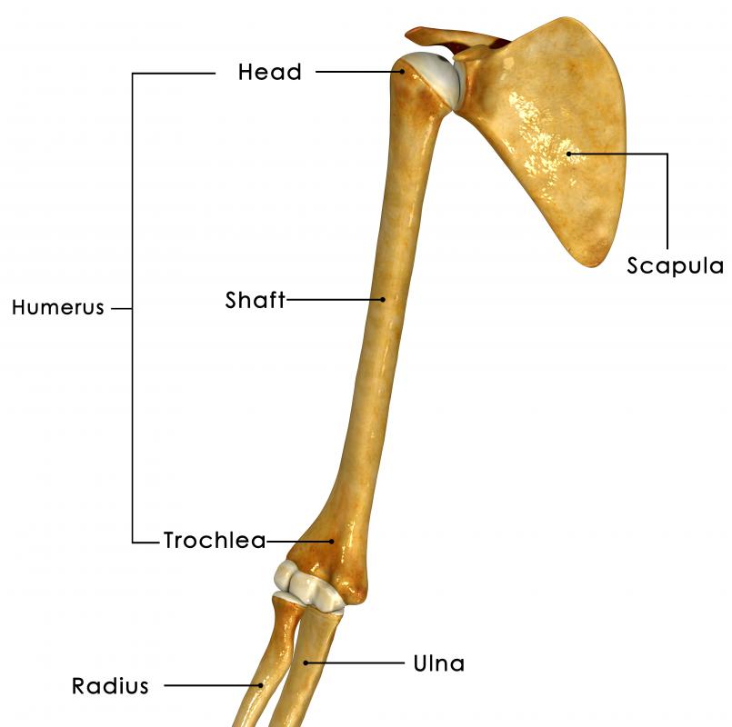
The upper arm bone, called the humerus, is articulated at its anterior end by a large, nearly perfectly hemispherical cap simply called the head. It nestles perfectly within a concavity of the scapula bone, or shoulder blade. This glenoid cavity and the caput humeri constitute the ball-and-socket joint, cushioned to swivel freely by softer bone tissue called cartilage. The joint is held together in place with the aid of tendons and ligaments, two types of connective tissue.

One of the tendons attached to the top tip of the glenoid cavity socket connects the bicep muscle of the upper arm. Opposite, a tendon attaches the triceps muscle, and the two counteract to enable the movements called flexion and extension of the shoulder. The most prominent muscle of the shoulder is the deltoideus, whose triangular shape encapsulates the shoulder in front, back, and laterally. One of the deltoid muscle’s attachment location is the rear lower lip of the shoulder blade. Another point connects to the clavicle, while a third insertion point is a thick tendon reaching nearly halfway down the humerus bone. Additional muscles connecting the humerus to the scapula include the large triangular subscapularis, the supraspinatus and infraspinatus, and the teres major and minor muscles.

The various muscles working in combination enable the shoulder joint to function with an extraordinary range of motion. In addition to flexion and extension, abduction and adduction are the opposing movement of the arms away from and back toward the central vertical axis of the body. The anatomy of the shoulder also enables rotation around the central axis of the arm bone. Most characteristic, accounting most for the shoulder joint’s versatility, is circumduction, defined by the free range of movement within a conical space.

For their sheer bulk, the muscles of the shoulder can also exert considerable force in most of its kinesthetic directions. Primarily keeping the joint together under force of strenuous activity are the thick tendons connecting the muscles to their respective bones. Ligaments also assist in this regard, but their more critical function is to constrain the joint from exceeding its maximum movement range. Syndesmology is the study of the connective tissue of joints and their relation to joint movement.
Notable among the several major ligaments of the shoulder joint is the articular capsule that completely encases it and prevents the separation of the ball from its socket by no more than 1 inch (2.5 cm). A prominent cartilaginous structure under the articular capsule is the glenoidal labrun, which is an extension to the circumference of the socket cavity. This protects its bone edge and also grips the ball. Though relatively small in size, several bursae — sacs mostly located where tendons contact bones containing synovia — contain a viscous fluid that is important in lubricating movement. Finally, though conventionally not included as part of the anatomy of the shoulder, are the blood vessels, lymph vessels, nerve fibers, and other tissue which support its healthy functioning.
AS FEATURED ON:
AS FEATURED ON:

-
![An anatomical illustration showing many muscles in the upper body and shoulder.]() By: mikiradicAn anatomical illustration showing many muscles in the upper body and shoulder.
By: mikiradicAn anatomical illustration showing many muscles in the upper body and shoulder. -
![A separated shoulder may occur when the ligament holding the collarbone and the shoulder together is damaged.]() By: Dr CanoA separated shoulder may occur when the ligament holding the collarbone and the shoulder together is damaged.
By: Dr CanoA separated shoulder may occur when the ligament holding the collarbone and the shoulder together is damaged. -
![The scapula bone is also known as the shoulder blade.]() By: 7activestudioThe scapula bone is also known as the shoulder blade.
By: 7activestudioThe scapula bone is also known as the shoulder blade.


Discuss this Article
Post your comments