At WiseGEEK, we're committed to delivering accurate, trustworthy information. Our expert-authored content is rigorously fact-checked and sourced from credible authorities. Discover how we uphold the highest standards in providing you with reliable knowledge.
What Is the Superior Semicircular Canal?
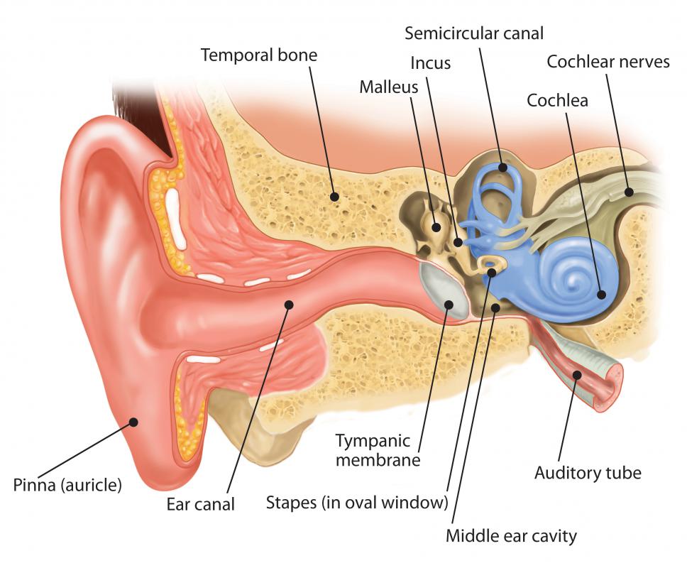
The superior semicircular canal is one of the three semicircular canals found in the vestibular apparatus, an organ in the inner ear that is responsible for the sense of equilibrium or balance. Also called the anterior semicircular canal, the superior semicircular canal is oriented at right angles to the posterior semicircular canal and the lateral semicircular canal. Together with the two other semicircular canals, the superior semicircular canal is responsible for detecting rotational or angular acceleration, and helps maintain balance and visual tracking when the head is turned.
Both the semicircular canals and the otolith organs belong to the vestibular system, and are situated inside a tubelike structure called membranous labyrinth. Endolymph, a fluid that is similar to the fluid within cells or intracellular fluid, can be found within this structure. The otolith organs, which include the utricle and the saccule, are responsible for detecting linear acceleration. Like the other two canals, the superior semicircular canal has a bulge or enlargement on one end called ampulla, which is where the receptors for equilibrium called sensory hair cells are located. These hair cells have about 20 to 50 hairlike extensions, are embedded in a gelatinous structure called cupula that helps increase the sensitivity of the hair cells to changes in equilibrium, and are also connected to sensory nerve fibers that transmit nerve signals to the eighth cranial nerve or the vestibulocochlear nerve.

When there is a rotational change in equilibrium, such as when the head is turned or when an impact forces the head to move sideways, the endolymph within the semicircular canal causes the cupula to move. The hair cells bend in a direction opposite to that of the rotational force. For instance, if the head rotates to the left, the endolymph causes the cupula to bend to the right, resulting in the stimulation of the hair cells.

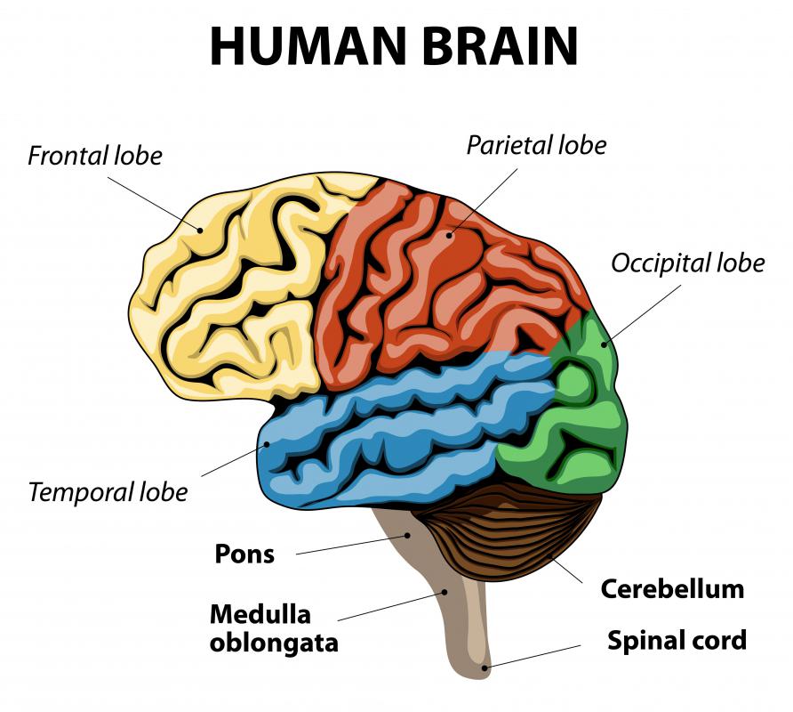
Once the stimulation of hair cells occurs, action potentials are generated and the sensory neurons of the eighth cranial nerve are activated. These neurons then transmit nerve signals to the cerebellum, the brain stem, and the spinal cord. The brain stem, particularly the part called oculomotor center, responds by controlling eye movements, while the spinal cord responds by stimulating the head, neck, and limb movements. The coordination of these different organs is needed to maintain and allow visual tracking, such as what occurs when the head is turned while reading or focusing on an object.
AS FEATURED ON:
AS FEATURED ON:

-
![There are three semicircular canals found in the vestibular apparatus, an organ in the inner ear.]() By: kocakayaaliThere are three semicircular canals found in the vestibular apparatus, an organ in the inner ear.
By: kocakayaaliThere are three semicircular canals found in the vestibular apparatus, an organ in the inner ear. -
![Once the stimulation of hair cells occurs, action potentials are generated and the sensory neurons of the eighth cranial nerve are activated.]() By: arkelaOnce the stimulation of hair cells occurs, action potentials are generated and the sensory neurons of the eighth cranial nerve are activated.
By: arkelaOnce the stimulation of hair cells occurs, action potentials are generated and the sensory neurons of the eighth cranial nerve are activated. -
![Sensory neurons transmit nerve signals to the cerebellum, the brain stem, and the spinal cord.]() By: designuaSensory neurons transmit nerve signals to the cerebellum, the brain stem, and the spinal cord.
By: designuaSensory neurons transmit nerve signals to the cerebellum, the brain stem, and the spinal cord. -
![Once the stimulation of hair cells occurs in the inner ear, sensory neurons of the eighth cranial nerve are activated, transmitting signals to the cerebellum, the brain stem and the spinal cord.]() By: designuaOnce the stimulation of hair cells occurs in the inner ear, sensory neurons of the eighth cranial nerve are activated, transmitting signals to the cerebellum, the brain stem and the spinal cord.
By: designuaOnce the stimulation of hair cells occurs in the inner ear, sensory neurons of the eighth cranial nerve are activated, transmitting signals to the cerebellum, the brain stem and the spinal cord.


Discuss this Article
Post your comments