At WiseGEEK, we're committed to delivering accurate, trustworthy information. Our expert-authored content is rigorously fact-checked and sourced from credible authorities. Discover how we uphold the highest standards in providing you with reliable knowledge.
What is the Process for Differential Diagnosis of Pleural Effusion?
A pleural effusion occurs when fluid collects in the area between the lung and the pleura, a membrane that is found between the lung and the chest cavity. This fluid buildup can be caused by a number of different disease processes. Detection of a pleural effusion, either by physical exam or radiograph, necessitates an investigation into the cause of the effusion. The most important aspect of forming a differential diagnosis of pleural effusion is performing a thoracentesis and obtaining a sample of the pleural fluid. Analysis of this fluid yields a large amount of information regarding the cause.
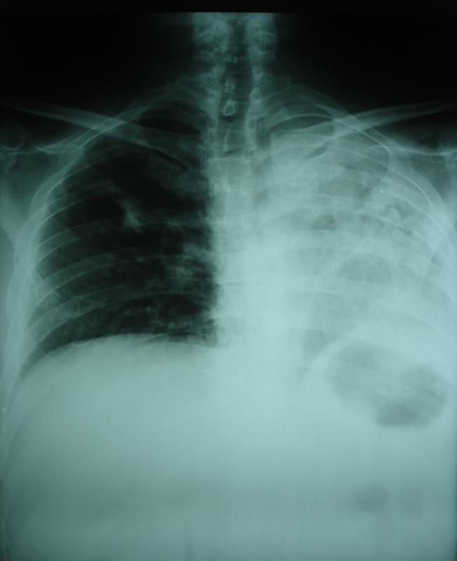
Pleural effusions can be diagnosed on the basis of clinical history, physical exam, and x-ray findings. Patients might report symptoms such as shortness of breath or pain on deep inspiration. On physical exam, doctors might identify an area of decreased resonance to percussion or a region of decreased breath sounds over the pleural effusion. Findings on chest x-ray can include blunting of the costophrenic angles formed by the meeting of the ribs and diaphragm, and areas of opacity within the lung fields.

After identifying its presence, the next step in the differential diagnosis of pleural effusion is performing a procedure called a thoracentesis. The importance of the thoracentesis cannot be minimized; in fact, doctors are taught to do this as soon as possible in cases of pleural effusion. With this procedure, a sterile needle is inserted between the ribs in order to obtain a sample of the fluid. The procedure can be done with the help of an ultrasound machine, or can be performed using physical exam maneuvers to localize the effusion.

The pleural fluid obtained by thoracentesis is sent to the laboratory for a number of tests. The first step in diagnosis rests on determining whether the fluid is an exudate or a transudate. Light’s critera are traditionally used to differentiate exudates from transudates. Pleural effusions are considered to be exudates if the ratio between the pleural fluid protein to the serum protein concentration is greater than 0.5. Additionally, if the pleural fluid lactate dehydrogenase (LDH) is greater than two-thirds the upper limit of normal, or if the ratio of pleural fluid LDH to serum LDH is greater than 0.6, the pleural effusion is considered to be an exudate.
Knowing whether the pleural effusion is exudative or transudative is important for diagnosis. Transudative pleural effusions are caused by imbalances in pressures within the chest cavity. Examples of causes of transudative pleural effusions include congestive heart failure, nephrotic syndrome, and hypoalbuminemia. In contrast, exudative pleural effusions are more commonly caused by infectious or inflammatory states. Examples of causes of exudative pleural effusions include pneumonia, tuberculosis, cancer, and connective tissue disorders.
There are other ways in which pleural fluid can be helpful in the differential diagnosis of pleural effusion. The fluid is often cultured to see if any bacterial species can be grown. It can be sent for cytogenetic analysis to see if there is any evidence of malignancy. High levels of amylase in the fluid can suggest pancreatitis, esophageal rupture, or cancer. Very low levels of glucose could indicate tuberculosis, lupus, or rheumatoid arthritis.
AS FEATURED ON:
AS FEATURED ON:

-
![A chest x-ray may help confirm the diagnosis of pleural effusion.]() By: Rob hyronsA chest x-ray may help confirm the diagnosis of pleural effusion.
By: Rob hyronsA chest x-ray may help confirm the diagnosis of pleural effusion. -
![A pleural effusion occurs when excess fluid builds around the lungs.]() By: Dr CanoA pleural effusion occurs when excess fluid builds around the lungs.
By: Dr CanoA pleural effusion occurs when excess fluid builds around the lungs.

Discuss this Article
Post your comments