At WiseGEEK, we're committed to delivering accurate, trustworthy information. Our expert-authored content is rigorously fact-checked and sourced from credible authorities. Discover how we uphold the highest standards in providing you with reliable knowledge.
What Is the Corneal Endothelium?
The corneal endothelium is a single thin layer of cells lining the interior, rear surface of the cornea in the eye. It is thus named after its location as well as the type of cells that form it, called endothelial cells; these cells are flat and are responsible for keeping the cornea clear. The corneal endothelium, also known as the posterior epithelium, faces the anterior chamber of the eye, which is located between the cornea and the part of the eye that it covers, called the iris.
Formation of the corneal endothelium takes place before birth, during the embryonic stage. Its origin is the neural crest, a group of cells that leave the neural tube to form a variety of cells — from melanin-producing cells, or melanocytes, in the skin to neurons in the nervous system. Upon birth, the endothelial cellular count is usually around 300,000 per cornea. By adulthood, however, the cell density decreases until it settles to a range of approximately 2,000 to 3,200 cells per square millimeter in each eye. The layer is usually composed of evenly sized cells, which form a hexagonal shape.

The anterior chamber, which the corneal endothelium faces, is located between the cornea and the iris. This space is filled with a thick, watery fluid called aqueous humor. This substance carries out several functions, which include inflating the eyeball and providing the eye with nutrition.
The back of the corneal endothelium has direct contact with the fluid in the anterior chamber. This positioning enables the layer of cells to transport necessary nutrients from the aqueous humor to the areas of the cornea that need it. At the same time, the corneal endothelium takes water from the corneal stroma — the part of the eye at the corneal endothelium's front border that strengthens the cornea — and transports it to the aqueous humor. Supporting the corneal endothelium at its anterior border is Descemet's membrane, which is also categorized as a "basement membrane" because it underlies this layer of cells.

Risk of the corneal endothelium failing or decreasing in density increases as people age or when they experience optical trauma. An example of a disease affecting the cellular layer is Fuchs' dystrophy, or Fuchs' endothelial dystrophy. This degenerative corneal disorder involves the thickening of Descemet's membrane and an accumulation of fluid in the endothelium, with visual impairment as a consequence. The layer can also be adversely affected by iritis, which is an inflammation of the iris, and glaucoma, which is characterized by optic nerve damage. Although various surgical techniques exist for treatment of such diseases, there is no method to repair the corneal endothelium itself.
AS FEATURED ON:
AS FEATURED ON:

-
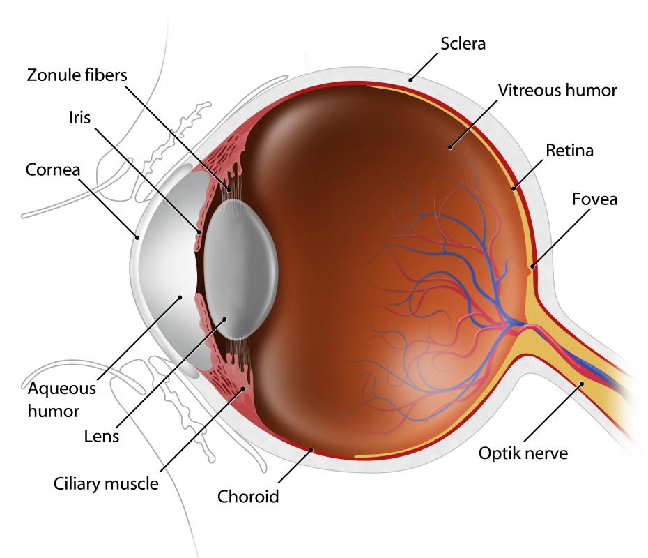
![The corneal endothelium is located between the cornea and the iris.]() By: Subbotina AnnaThe corneal endothelium is located between the cornea and the iris.
By: Subbotina AnnaThe corneal endothelium is located between the cornea and the iris. -
![The corneal endothelium cells are responsible for keeping the cornea clear, allowing light into the eye.]() By: kocakayaaliThe corneal endothelium cells are responsible for keeping the cornea clear, allowing light into the eye.
By: kocakayaaliThe corneal endothelium cells are responsible for keeping the cornea clear, allowing light into the eye.

Discuss this Article
Post your comments