At WiseGEEK, we're committed to delivering accurate, trustworthy information. Our expert-authored content is rigorously fact-checked and sourced from credible authorities. Discover how we uphold the highest standards in providing you with reliable knowledge.
What Is the Choroid Fissure?
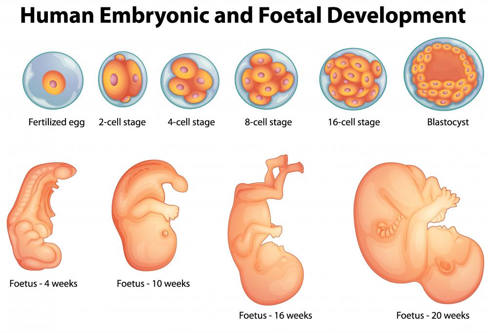
The choroid fissure is an optical component that is involved in early fetal development. This tubular structure acts as a transit route for optic nerves exiting the eye and entering blood vessels. A properly developing choroid fissure will fuse together during development. Failure to fuse can cause a hole, called a coloboma, within the eye.
The eye begins to develop about four weeks into fetal development. This begins when embryonic cells create two recesses in the forebrain. Growth expansion of these recesses causes slim structures called optic stalks to begin to form. Optic stalks connect the recesses — later called optic vesicles and then optic cups — to the brain wall and eventually become the optic nerve.

Running beneath the optic stalks is the choroid fissure. The fissure contains the hyroid artery and the hyroid vein. Nutrient blood vessels travel through these structures into the forming lens. When the fissure fuses closed, the vein and artery are trapped inside the optic stalks. They then develop into the retinal artery and vein.
Creation of the visual system happens quickly during embryonic development. The choroid fissure should be fused shut at the sixth week of gestation. This process happens very quickly, so it is imperative that every part of the process operates properly. When a choroid fissure doesn't fuse, a coloboma forms.

Colobomas can appear in one or both eyes. The severity of vision loss depends on the size of the coloboma and its location. Coloboma iridis, the most common form, affects the iris of the eye. This simpler defect results in less loss of visual accuity than other colobomas. Light sensitivity can be increased with this form.
A coloboma also can form in the fundus of the eye. The fundus contains optic components that include the retina, macula, optic disc and a layer of blood vessels and tissue called the choroid. If the choroid fissure is mostly closed, then the coloboma will occur in the front of the fundus and cause fewer problems than the alternative. A more open choroid fissure will extend the coloboma back into the fundus and cause issues with central vision.
Associated eye problems such as microphthalmos, or underdevelopment of the eyeball, can exacerbate this vision loss. There is no cure for colobomas. Young children might be monitored by a vision specialist to diagnose and monitor the progression of the condition.
AS FEATURED ON:
AS FEATURED ON:

-
![Light sensitivity may occur as a result of coloboma iridis.]() By: petarpaunchevLight sensitivity may occur as a result of coloboma iridis.
By: petarpaunchevLight sensitivity may occur as a result of coloboma iridis. -
![The choroid fissure is an optical component that is involved in early fetal development.]() By: blueringmediaThe choroid fissure is an optical component that is involved in early fetal development.
By: blueringmediaThe choroid fissure is an optical component that is involved in early fetal development.

Discuss this Article
Post your comments