At WiseGEEK, we're committed to delivering accurate, trustworthy information. Our expert-authored content is rigorously fact-checked and sourced from credible authorities. Discover how we uphold the highest standards in providing you with reliable knowledge.
What is the Anatomy of the Knee?
The anatomy of the knee includes a number of structures related to its function, the movements of flexion and extension. These include the three bones of the knee joint — the femur, tibia, and patella — several attaching muscles and tendons, the ligaments joining the bones together, and the structures contained within the joint itself. A type of synovial joint called a hinge joint, the articulation at the knee allows movement in a front-to-back direction only.
The anatomy of the knee, from the shape of the bones to the muscles that cross the joint, is what makes this movement possible. Meeting at the knee are the femur bone of the thigh, the tibia bone of the shin, and the patella bone of the kneecap. The knee joint is found beneath the patella and between the femur and tibia.

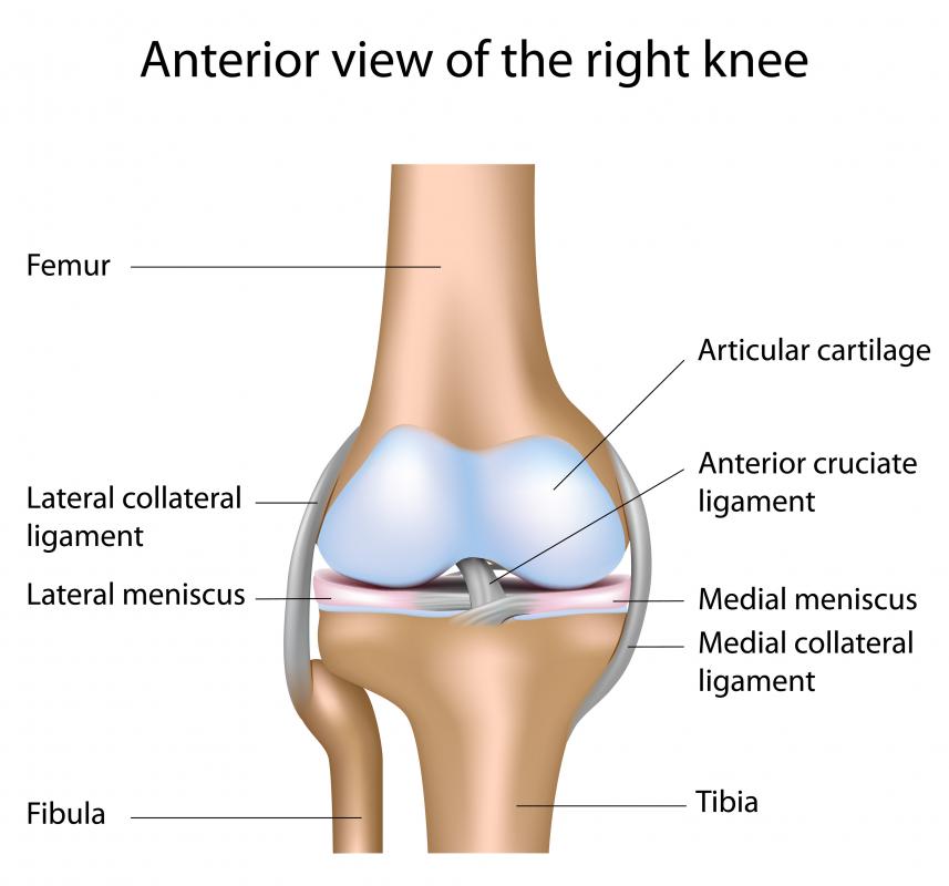
Several ligaments connect these bones and protect the knee against excessive or unstable forces. The extracapsular ligaments include the patellar ligament, which links the inferior or lower surface of the patella to the tibia below. The medial and lateral collateral ligaments run vertically along the inside and outside of the knee, respectively. Finally, two popliteal ligaments are on the back side of the knee.

Beneath the patella and inside the joint capsule are the intracapsular ligaments, which include the anterior and posterior cruciate ligaments crossing the joint like an X. This area also includes the transverse ligament, which links the medial and lateral menisci horizontally. Several smaller ligaments also can be found here.
Also relevant to the anatomy of the knee are several muscles of flexion and extension. The quadriceps is the collective name for the large group of muscles in the anterior thigh that end in a tendon just above the patella. This tendon’s fibers cross the top of the patella bone and form the patellar ligament below. They are the muscles responsible for extension or straightening of the knee joint. On the back of the thigh is a group of muscles called the hamstrings that cross the back of the knee. They cause knee flexion or bending by pulling upward on the back of the lower leg during muscle contraction.
The anatomy of the knee would not allow for extension, flexion, or absorption against impact forces were it not for the hinge joint within. Surrounded by a lining called a synovial membrane, the joint capsule contains the adjoining ends of the femur and tibia bones, synovial fluid to lubricate the joint against friction, cartilaginous disks called menisci to cushion the bones and absorb impact, and the intracapsular ligaments. The anatomy of the knee joint not only keeps the bones from rubbing against one another but also resists the wearing down of the joint’s cartilage over time.
AS FEATURED ON:
AS FEATURED ON:

-
![An anterior view of the right knee.]() By: AlilaAn anterior view of the right knee.
By: AlilaAn anterior view of the right knee. -
![The anatomy of the knee includes a number of structures related to the flexion and extension of the knee.]() By: DirimaThe anatomy of the knee includes a number of structures related to the flexion and extension of the knee.
By: DirimaThe anatomy of the knee includes a number of structures related to the flexion and extension of the knee.

Discuss this Article
Post your comments