At WiseGEEK, we're committed to delivering accurate, trustworthy information. Our expert-authored content is rigorously fact-checked and sourced from credible authorities. Discover how we uphold the highest standards in providing you with reliable knowledge.
What is the Anatomy of the Ankle?
The anatomy of the ankle includes of all structures contained in and surrounding the ankle, or talocrural, joint. These include the contents of the joint capsule such as the ends of the articulating bones, joint cartilage, and synovial fluid. Ankle anatomy also refers to the ligaments enclosing the capsule and holding the bones together, the muscles and tendons crossing the ankle joint, and the fat and skin around it.
A synovial joint, the ankle can produce the hinging movements of dorsiflexion and plantarflexion. It is capable of absorbing nearly the entirety of the body’s weight during standing, walking, running, and jumping movements.

Like any of the body’s movable joints, the talocrural joint is a junction of two or more bones, specifically the tibia and fibula bones in the lower leg with the talus bone of the ankle. Between these shin bones and the talus below is the synovial capsule or joint space. Within this lubricated, fluid-filled cavity is cartilage to cushion the bones against each other during weight-bearing movements as well as the front-to-back hinging movements of dorsiflexion and plantarflexion. Dorsiflexion is the act of lifting the top or dorsal surface of the foot upward toward the shin, while plantarflexion is the act of pressing the bottom or plantar surface of the foot downward away from the shin.

The anatomy of the ankle also includes two more articulations — the inferior tibiofibular joint above and the subtalar joint below. The inferior tibiofibular joint is where the lower ends of the tibia and fibula meet immediately above the ankle joint. A type of joint known as a syndesmosis that is held together by an interosseous ligament, its bones are permitted very little movement against one another. Below the talocrural joint on the underside of the talus bone is the subtalar joint.
Found where the talus meets the top surface of the calcaneus or heel bone in the foot, the subtalar is the synovial articulation that allows the movements of eversion and inversion. This is the rolling of the ankle from side to side. Together with dorsiflexion and plantarflexion, these movements make circling the ankle possible and therefore the subtalar joint can be incorporated into an understanding of the anatomy of the ankle.
All of the bones at all of the ankle’s joints are held together by strong ligaments, another important component of the anatomy of the ankle. The tibia and fibula are joined by their single interosseous ligament as well as the anterior and posterior tibiofibular ligaments in front and behind, respectively, while each bone has its own ligaments connecting it to the talus. The medial malleolus of the tibia, the rounded bony prominence felt on the inside of the ankle, is joined to both the talus and the heel bone by the broad deltoid ligament. Likewise, the lateral malleolus of the fibula, a similar bony prominence felt on the outside of the ankle, is linked to the talus via the anterior and posterior talofibular ligaments and to the calcaneus via the calcaneofibular ligament.
A discussion of the anatomy of the ankle would not be complete without including the major muscles that act on the ankle joint. The large muscles of the calf on the posterior lower leg, the gastrocnemius and soleus, are responsible for the downward-hinging motion of plantarflexion, since they cross the back of the ankle as the Achilles tendon and attach to the heel bone. Dorsiflexion is initiated by several muscles of the anterior lower leg or shin that cross the ankle joint as individual tendons and insert in the foot, including the tibialis anterior, extensor digitorum longus, and extensor hallucis longus.
AS FEATURED ON:
AS FEATURED ON:

-
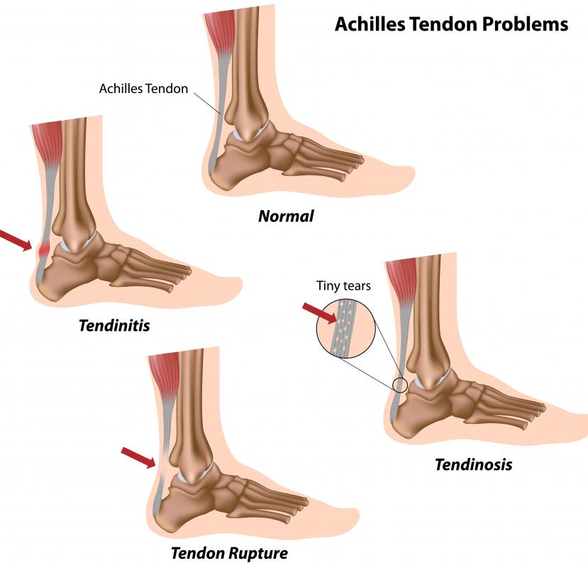
![This diagram shows the parts of the ankle, along with some common problems with the Achilles tendon.]() By: AlilaThis diagram shows the parts of the ankle, along with some common problems with the Achilles tendon.
By: AlilaThis diagram shows the parts of the ankle, along with some common problems with the Achilles tendon. -
![The location where the talus bone in the foot meets with the bottom of the tibia and fibula is known as the ankle joint.]() By: andreaxtThe location where the talus bone in the foot meets with the bottom of the tibia and fibula is known as the ankle joint.
By: andreaxtThe location where the talus bone in the foot meets with the bottom of the tibia and fibula is known as the ankle joint.

Discuss this Article
Post your comments