At WiseGEEK, we're committed to delivering accurate, trustworthy information. Our expert-authored content is rigorously fact-checked and sourced from credible authorities. Discover how we uphold the highest standards in providing you with reliable knowledge.
What is Doppler Ultrasonography?
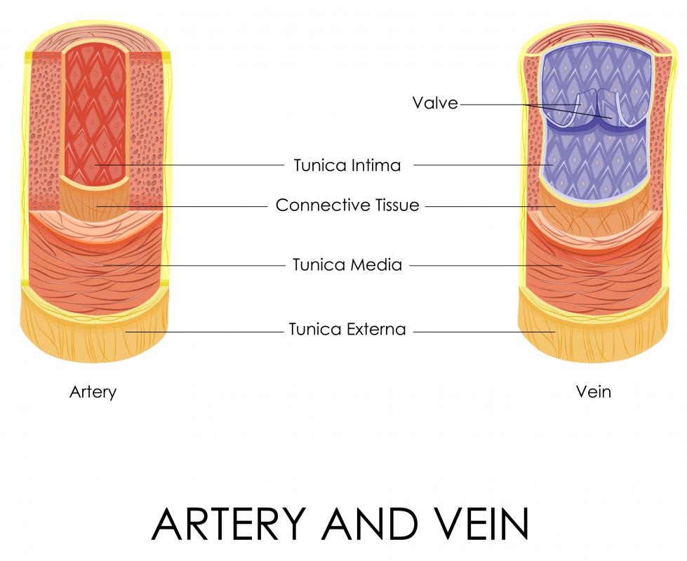
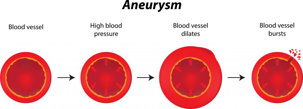
Doppler ultrasonography, also known as Doppler ultrasound, is an imaging technique that provides visualization on the flow of blood through the arteries. It is a non-invasive and painless procedure that uses sound waves to diagnose a disease. The procedure may be performed as an option to x-ray imaging techniques, such as venography and arteriography, which entail dye injection into the blood vessel to make it clearly visible on x-ray. Doppler ultrasonography can help spot blood clots, aneurysms, and heart valve problems, among others. It can be carried out in major veins and arteries of the body such as the legs, neck, and arms.
There are basically three forms of Doppler ultrasonography: color, power, and spectral. With the aid of a computer, color Doppler translates Doppler metrics into a range of colors to give a picture of the direction and flow rate of blood through an artery. Power Doppler, can provide more blood flow information, especially in situations where there is nominal blood flow; it is usually employed in the assessment of blood flow through arteries located in solid organs. Spectral Doppler displays blood flow details in a graph rather than in picture form.

Doppler ultrasonography uses two complementary principles — ultrasound and Doppler. Ultrasound is based on the principle that high-frequency sound will be bounced back from its target to where it came from. The Doppler principle, on the other hand, holds that that the pitch of a sound heightens as the source of the sound comes nearer to the person listening to it; conversely, the pitch of the sound decreases as the source of the sound moves away from the listener. Applying the two principles leads to the formation of an image of the organ being examined using Doppler ultrasonography.

A technician, known as a sonographer, performs the Doppler ultrasonography. She uses a hand-held transducer, also known as a probe. She presses the transducer on the skin of the person in the area where the organ being checked is located. Gel is often applied on the area of the skin to be probed for easier movement of the transducer. The image of the organ is captured at different angles in an x-ray film, disc, or in paper for use for the doctor who requested for the Doppler ultrasonography.
No preparation is needed prior to the conduct of Doppler ultrasonography. It may be done as an outpatient procedure, and does not require post-procedure care.
AS FEATURED ON:
AS FEATURED ON:

-
![Ultrasonography uses ultrasonic waves to form images of a patient's internal organs.]() By: Zsolnai GergelyUltrasonography uses ultrasonic waves to form images of a patient's internal organs.
By: Zsolnai GergelyUltrasonography uses ultrasonic waves to form images of a patient's internal organs. -
![Doppler ultrasonography is an imaging technique that provides visuals of the flow of blood through the arteries and veins.]() By: stockshoppeDoppler ultrasonography is an imaging technique that provides visuals of the flow of blood through the arteries and veins.
By: stockshoppeDoppler ultrasonography is an imaging technique that provides visuals of the flow of blood through the arteries and veins. -
![The Doppler ultrasound machine uses the Doppler effect to create a moving image of blood flow in the body.]() By: zayacskThe Doppler ultrasound machine uses the Doppler effect to create a moving image of blood flow in the body.
By: zayacskThe Doppler ultrasound machine uses the Doppler effect to create a moving image of blood flow in the body. -
![A blocked artery in the heart may be detected using a medical Doppler ultrasound device.]() By: Alexandr MitiucA blocked artery in the heart may be detected using a medical Doppler ultrasound device.
By: Alexandr MitiucA blocked artery in the heart may be detected using a medical Doppler ultrasound device. -
![An ultra sound can be used to detect aneurysms.]() By: joshyaAn ultra sound can be used to detect aneurysms.
By: joshyaAn ultra sound can be used to detect aneurysms. -
![A doppler ultrasound may be used to check for blockages in the arteries and veins in the neck.]() By: Dario Lo PrestiA doppler ultrasound may be used to check for blockages in the arteries and veins in the neck.
By: Dario Lo PrestiA doppler ultrasound may be used to check for blockages in the arteries and veins in the neck. -
![Doppler ultrasonography may be used to detect heart problems.]() By: Vladislav GajicDoppler ultrasonography may be used to detect heart problems.
By: Vladislav GajicDoppler ultrasonography may be used to detect heart problems.


Discuss this Article
Post your comments