At WiseGEEK, we're committed to delivering accurate, trustworthy information. Our expert-authored content is rigorously fact-checked and sourced from credible authorities. Discover how we uphold the highest standards in providing you with reliable knowledge.
What Is Descemet's Membrane?
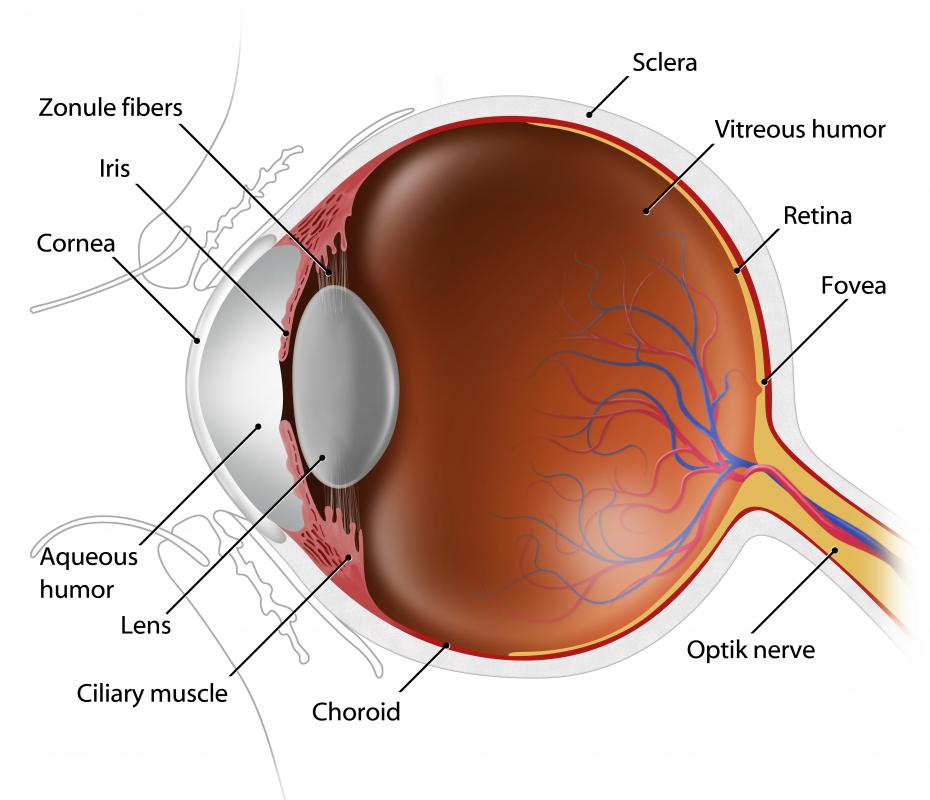
Named after the French physician, Jean Descemet, Descemet's membrane is an elastic layer of membranous tissue that lies between the endothelial layer of the cornea and the corneal layer called the stroma. It functions as a protective barrier against eye injury and infection and is able to regenerate itself after an injury. Descemet's membrane lies beneath the epithelial tissue of the eye, making it a basement membrane, and is composed of collagen produced by the endothelial cells that lay beneath it.
The cornea is the transparent outer layer of the eye, which is comprised of five layers of tissue. The first layer is called the epithelium. Epithelial tissue, nervous tissue, muscle tissue and connective tissue make up the four categories of animal tissue.

In both animals and humans, the epithelium lines body cavities, forms some of the body's glands, and lines the body's internal and external surfaces. It can also regenerate itself after injury. The epithelium performs two functions: to keep out foreign matter such as dirt and germs, and to absorb oxygen and nutrients and then distribute them to other parts of the cornea.

The second layer of the cornea is known by several names: Bowman's layer or Bowman's membrane, the anterior limiting lamina, and the anterior elastic lamina. Bowman's membrane is a transparent stratum of layered protein fibers, or collagen, that lays directly beneath the epithelium. Beneath the Bowman's layer is the stroma, which is by far the thickest part of the cornea, comprising about ninety percent of the cornea's density.
The fourth layer of the cornea, located under the stroma, is Descemet's membrane, which is a thin, transparent, elastic, and very strong sheet of tissue. It is known by several other names: the posterior limiting lamina, the posterior elastic lamina, the lamina elastica posterior, and the membrane of Demours. The primary function of Descemet's membrane is to protect the inner cornea from invasive or harmful material.
A condition called Wilson's disease makes Descemet's membrane susceptible to abnormal amounts of copper deposits, which form a ring around the eye's iris, or the colored part of the eye. This is called the Kayser-Fleischer ring. The ring itself causes no symptoms and isn't always visible to the naked eye.
Below Descemet's membrane is the endothelium, which is the fifth and innermost layer of the cornea. This layer removes excess fluids from the cornea. Endothelial cells can't regenerate after injury or disease, and losing too many endothelial cells can lead to corneal edema, or too much fluid in the cornea, and eventual blindness.
AS FEATURED ON:
AS FEATURED ON:

-
![The cornea is the clear layer of tissue at the front of the eye.]() By: Subbotina AnnaThe cornea is the clear layer of tissue at the front of the eye.
By: Subbotina AnnaThe cornea is the clear layer of tissue at the front of the eye. -
![The cornea allows light to enter into the eye.]() By: kocakayaaliThe cornea allows light to enter into the eye.
By: kocakayaaliThe cornea allows light to enter into the eye.

Discuss this Article
Post your comments