At WiseGEEK, we're committed to delivering accurate, trustworthy information. Our expert-authored content is rigorously fact-checked and sourced from credible authorities. Discover how we uphold the highest standards in providing you with reliable knowledge.
What Is 3D Ultrasound?
The 3D ultrasound is a medical technique used for observing a fetus. Traditional 2D ultrasound can get a 3D view of a fetus by tilting around and observing different snapshots to see dimensions and position. Actual 3D ultrasound, however, uses computer programs to reconstruct a full three-dimensional representation of the fetus, which can then be explored fully.
A 3D ultrasound is acquired by emitting high-frequency sound waves, using a handheld probe to direct them. These sound waves reflect back, giving a good idea of the shape and position of the baby. The composite of many different images creates a surprisingly high-quality three dimensional image, which can be crucial in ensuring a doctor can see any developing problems that may be occurring. A diagnostic ultrasound may reveal any number of complications with the mother, including irregular bleeding, inconsistencies with the ovaries, or an irregular placement of the placenta. This ultrasound can also reveal a great deal about the baby, including its sex, how large it is, how far along in term it is, and whether it suffers from some early physical defects like a cleft palette.

More and more mothers are choosing to take advantage of 3D ultrasound for non-diagnostic reasons, outside of a hospital or clinic setting. Locations known as sonogram studios offer services where they will let a mother undertake an elective 3D version to get a better image of their baby. Many psychologists have noted that this can help prenatal bonding between the mother and the child, and for many couples it helps them bond over the birth to come.

Elective 3D ultrasound is usually one of a number of options offered at these sonogram studios, a step up from a basic 2D ultrasound, usually only used to determine the sex of the baby. This ultrasound package at an elective facility might cost around $300 US Dollars (USD), and include not only the in-studio ultrasound, but the creation of a DVD for home viewing, or prints of stills from the ultrasound. A more recent service allows ultrasounds to be put online behind a password, allowing friends and family around the world to view the baby.

Often movement is added to these images by combining many different ultrasound stills. In this case the result is usually called a 4D ultrasound, and these videos are what can be shared among friends or presented on a DVD. The 4D ultrasound does not have a great deal of extra diagnostic merit, but does give a more visceral feeling of the baby being alive, making it psychologically much more satisfying to many parents.
The history of 3D ultrasound goes back to the 1970s, when researchers in Scotland began working on a multiplanar scanner. As computers developed, a true 3D image became more feasible, and by the 1980s a number of working groups were developing their own prototypes. In 1984, the first concrete work on a Japanese 3D method began, and was successful by 1986. Through the 1990s, as computers developed and groups continued advancing the technology, the resolution and speed of 3D ultrasounds increased rapidly, and by the year 2000 the technology had reached near its current level.
AS FEATURED ON:
AS FEATURED ON:

-
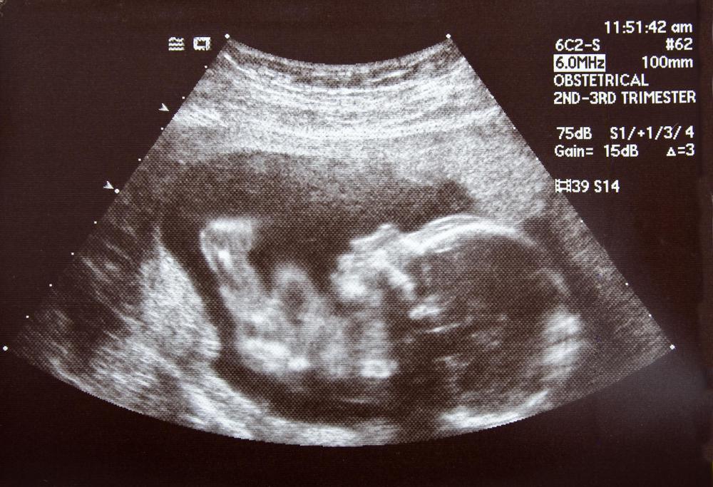
![A traditional 2D ultrasound may be moved and tilted to provide three-dimensional view.]() By: Darren BrodeA traditional 2D ultrasound may be moved and tilted to provide three-dimensional view.
By: Darren BrodeA traditional 2D ultrasound may be moved and tilted to provide three-dimensional view. -
![3D ultrasounds use computer programs to reconstruct a three-dimensional representation of a fetus in the womb.]() By: Giovanni Cancemi3D ultrasounds use computer programs to reconstruct a three-dimensional representation of a fetus in the womb.
By: Giovanni Cancemi3D ultrasounds use computer programs to reconstruct a three-dimensional representation of a fetus in the womb. -
![3D ultrasound uses the technology of a traditional ultrasound with the help of computer software to create better images.]() By: Monkey Business3D ultrasound uses the technology of a traditional ultrasound with the help of computer software to create better images.
By: Monkey Business3D ultrasound uses the technology of a traditional ultrasound with the help of computer software to create better images.


Discuss this Article
Post your comments