At WiseGEEK, we're committed to delivering accurate, trustworthy information. Our expert-authored content is rigorously fact-checked and sourced from credible authorities. Discover how we uphold the highest standards in providing you with reliable knowledge.
What are Superior Colliculi?
The superior colliculi are paired structures in the part of the brainstem called the tectum. They are located around the pineal gland, below the thalamus and above the inferior colliculi. These structures are located on the roof of the midbrain, and the Latin term for roof is tectum, so they are also called optic tectum. Both superior colliculi receive visual input originating from the retina and other sensory input from the inferior colliculi, spinal cord, cerebellum, pretectal region, periaqueductal gray, and substantia nigra. The function of the superior colliculi not only involves the direction of eye movements, but also the multimodal integration of acoustic and somatosensory information for attention and spatial orientation.
In neuroanatomy, the superior and inferior colliculi are known as corpora quadrigemina, which is a Latin term that means quadruplet bodies. Each superior colliculus is conventionally divided into seven layers. The three topmost layers are termed superficial layers, the next two are called intermediate layers, and the innermost two are called deep layers.

Included in the superficial layers are lamina I, II, and III, or stratum zonale, stratum griseum superficiale, and stratum opticum, respectively. Lamina III is also known as the optic layer because the axons of the optic nerve come together in this layer. The three superficial layers receive sensory input from the retina, pretectum, parabigeminal nucleus, and vision-related regions of the cerebral hemispheres, such as the primary visual cortex, visual cortex, and frontal eye fields.

The intermediate layers are divided into lamina IV and V, or stratum griseum intermediale and stratum album intermediale, respectively. Out of the seven layers, lamina IV is the thickest, and neuroanatomists frequently subdivide it further into the upper and lower parts. The deep layers include lamina VI and VII, or stratum griseum profundum and stratum album profundum, respectively. Both the intermediate and deep layers receive input from multiple association areas of the human brain.

In terms of output or efferents, both superior colliculi have axonal projections into various subcortical structures, such as the inferior colliculus, reticular formation, spinal cord, lateral geniculate nucleus, and pulvinar of the thalamus. The pulvinar is regarded as a center of image interpretation that helps the body maintain a stable visual environment despite changes in the position of the retina. Through their associations with various parts of the body that control spatial recognition and position, the superior colliculi are able to facilitate head and eye motion toward visual and auditory stimuli, and assist in rapid eye movements called saccades.
AS FEATURED ON:
AS FEATURED ON:

-
![The superior colliculi interpret visual cues while the inferior colliculi interpret auditory information.]() By: Alila Medical MediaThe superior colliculi interpret visual cues while the inferior colliculi interpret auditory information.
By: Alila Medical MediaThe superior colliculi interpret visual cues while the inferior colliculi interpret auditory information. -
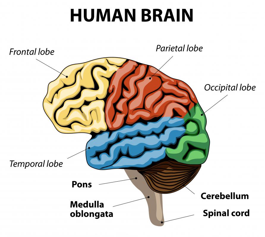
![The superior colliculi receive sensory input from the cerebellum.]() By: designuaThe superior colliculi receive sensory input from the cerebellum.
By: designuaThe superior colliculi receive sensory input from the cerebellum. -
![Both superior colliculi receive visual input originating from the retina.]() By: ikoBoth superior colliculi receive visual input originating from the retina.
By: ikoBoth superior colliculi receive visual input originating from the retina.


Discuss this Article
Post your comments